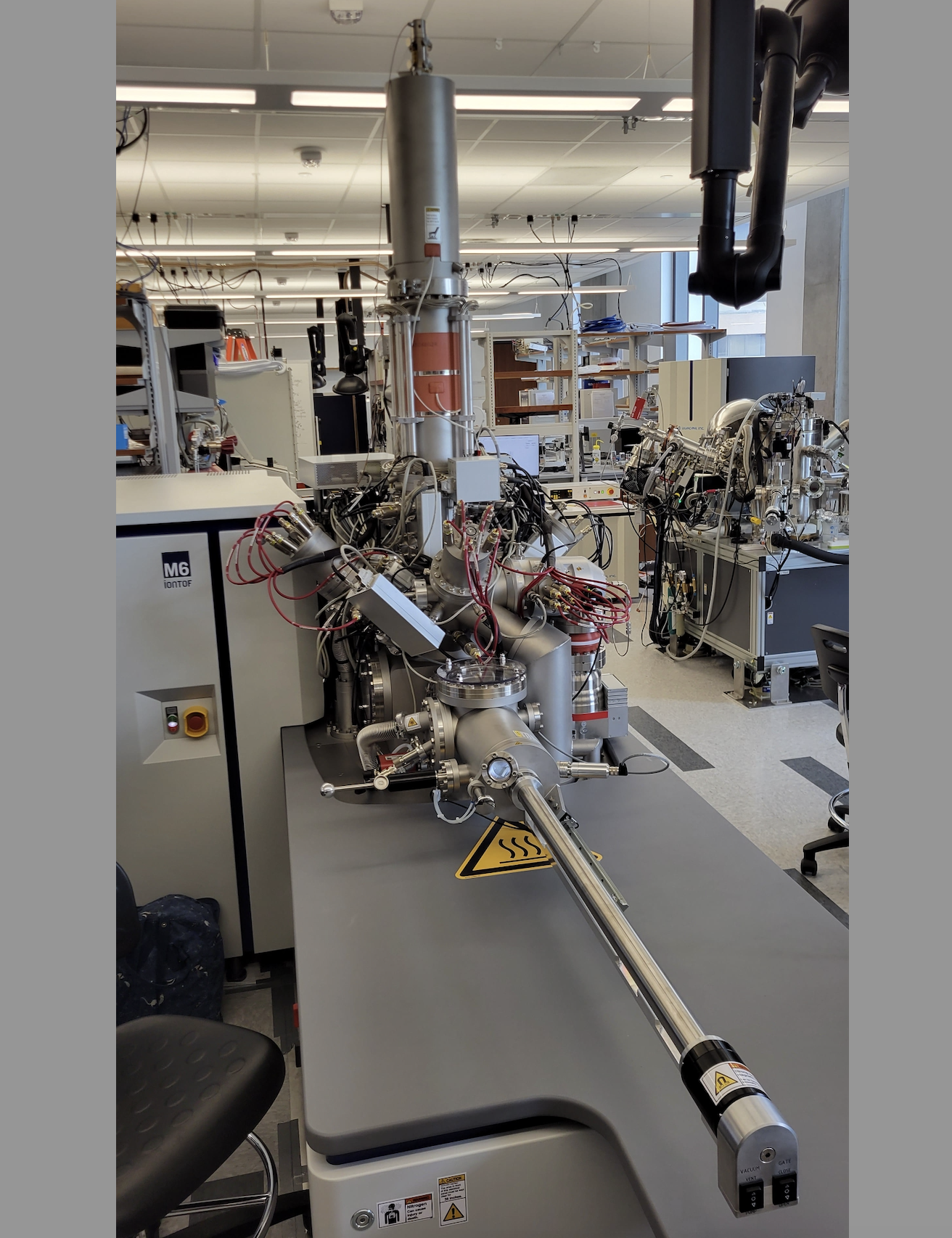
 Figure 1. The new ionTOF M6 TOF-SIMS instrument located in EER 6.636.
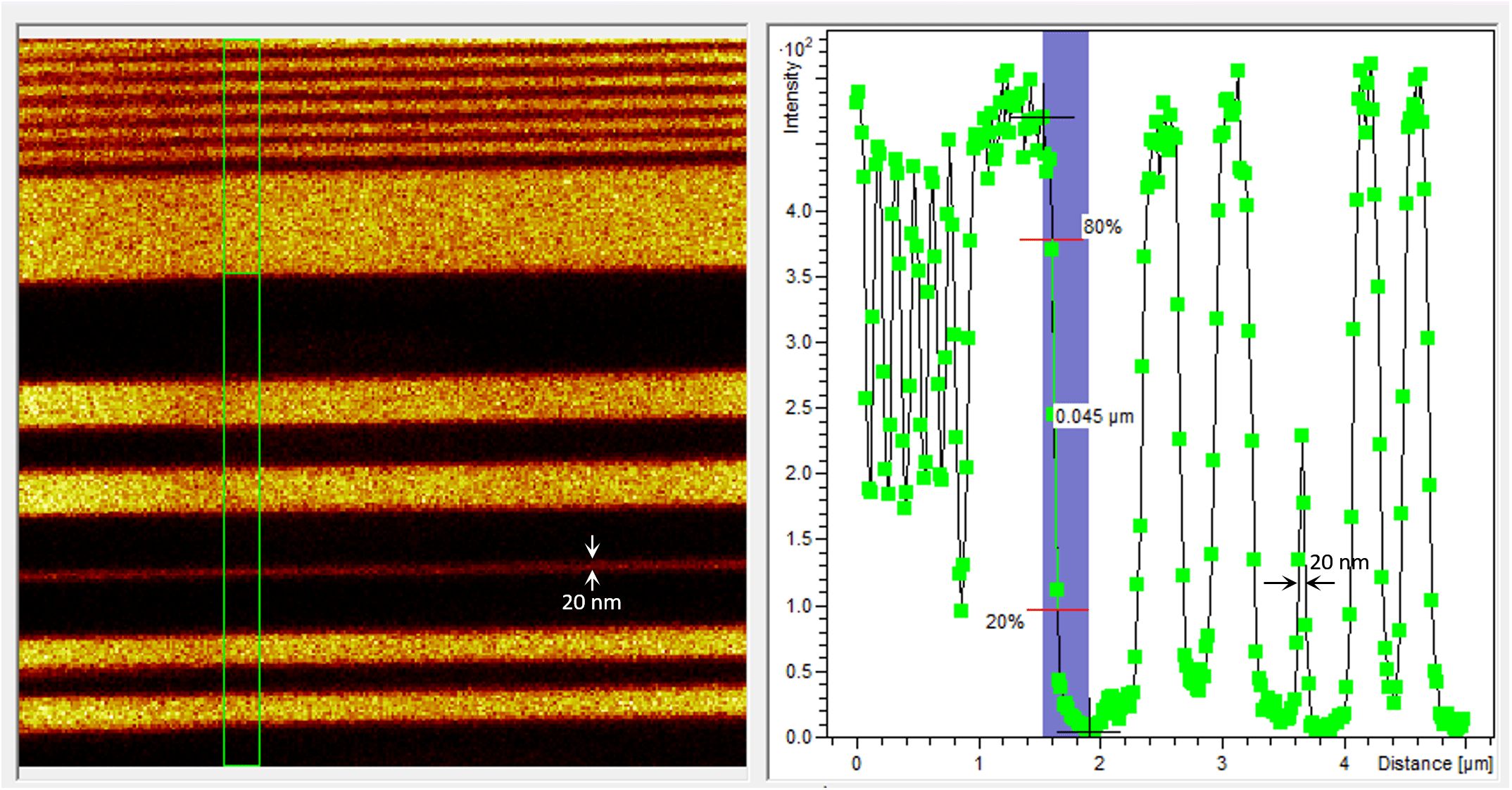
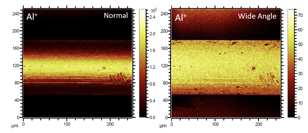
Highly versatile, the M6 is significantly advancing the capabilities of the old TOF.SIMS 5 ion microscope by demonstrating superior performance in all fields of SIMS applications. The lateral resolution is improved by a factor of 5 from ~ 100 nm (TOF.SIMS 5) to ~ 20 nm (M6) – Fig. 2; the mass resolution is improved by a factor of > 3 from ~10, 000 (TOF.SIMS 5) to > 30,000 (M6); the M6 also has > 3 times higher elemental sensitivity. Another impressive feature of the M6 is its unique delayed extraction mode for high transmission with simultaneous high lateral (< 50 nm) and high mass resolution (> 10,000), something that is unavailable on our old TOF.SIMS 5, but highly demanded by our users. With the addition of a focused ion beam (FIB) capability on the M6 instrument for 3D and cross-sectional analysis, extremely rough samples with voids and samples that exhibit strong local variations in density or sputter yield, such as batteries, fuel cells, pharmaceutical compounds, real-life photovoltaics, and microelectronic devices, etc., can be analyzed in cross section (Fig. 3). Such samples are almost impossible to directly analyze by conventional SIMS depth profiling. In conjunction with the cryogenic capability, the FIB capability will generate tremendous demand among our users since many of their samples are rough and hard to analyze by normal TOF-SIMS. Finally, the wide-angle secondary ion acceptance mode can provide high resolution TOF-SIMS maps of very rough features by increasing significantly the edge signals (Fig. 4).
Importantly, the greatly improved capabilities of the M6 system, such as unparalleled mass and spatial resolution, and elemental sensitivity, will enable a broad range of new science in key research areas in energy, electronics, healthcare, pharmaceuticals, etc. The new M6 instrument will provide the necessary means to perform advanced, 3D chemical analysis from nanometer to macroscopic level, enhancing the analytical options for the UT Austin research community and beyond.
 Figure 2. The M6 ion microscope is able to image aluminum lines 20 nm thick.

Figure 3. Cryo (~83 K) TOF-SIMS high lateral resolution imaging on a Li/Polymer battery cross section prepared by FIB inside the M6.

Figure 4. The wide-angle secondary ion acceptance capability on the M6 allows for edge observation in rough samples. Aluminum wire mapped in both normal and wide-angle high lateral resolution TOF-SIMS imaging modes.

